Introduction
Macrophages are a type of innate immunocyte that play an important role in immune homeostasis [1]. During the occurrence and development of pathogenic infection, macrophages act as scavengers to clear damaged tissue, participate in the inflammatory response and maintain immune balance through different functions, including phagocytosis, chemotaxis, polarization, adherence and secretion [2]. The recognition of damage-associated molecular patterns (DAMPs) or nutrient-based signals by several types of regulatory receptors, such as Toll-like receptors (TLRs), RIG-I-like receptors (RLRs), NOD-like receptors (NLRs) and lipid nuclear receptors (PPARγ, ERRγ), has been reported to trigger macrophage metabolic reprogramming [3-8].
Musculin (MSC), also known as myogenic repressor (MyoR) or activated B cell factor-1 (ABF-1), is a basic helix-loop-helix transcription repressor. Historically, the function of MSC has been studied in the context of mammalian skeletal myogenesis, tissue development, cell differentiation, and tissue regeneration [9]. Studies have shown that MSC is expressed not only [10] in organs and tissues of non-muscle lineages (including the adult brain, heart, liver, lung, small intestine, tonsils, etc.), but also [11] in immune organs and B cells of the spleen, thymus and lymph nodes, especially in adult lymphoid tissues (including the lymph nodes, appendix, and bone marrow) [12]. MSC was first identified in mouse skeletal precursors [13-15], and was recently found in mouse T cells. Moreover, MSC plays an important role in the development and transdifferentiation of regulatory T cells (Tregs) [16]. MSC expression in induced Tregs (iTregs) generated from naive T cells (Th0) increases in the presence of transforming growth factor β (TGF-β), and MSC deficiency can reduce Foxp3 expression and impair the ability of iTregs to inhibit the Th2 response, leading to the transdifferentiation of iTregs into Th2 cells. In addition, MSC promotes iTreg development by silencing Th2-related genes.
Our previous studies reported that MSC is highly associated with inflammatory cytokines and pathological processes [15]. MSC influences the differentiation of helper T cell (Th) subsets, especially Treg subsets. Additionally, MSC is highly expressed in alloantigen stimulated Th17 cells but not in iTreg subsets; a lack of MSC can lead to severe inflammation in murine colitis; and MSC can regulate the expression of IL-22 in innate lymphoid cells (ILCs) [17, 18]. Although MSC is closely associated with various types of immunocytes, its role in macrophages remains unclear.
The aim of this investigation was to explore the regulatory role of MSC in macrophages ex vivo under proinflammatory conditions. MSC knockout (MSC–/–) mice were used as experimental animals, and peritoneal primary macrophages were obtained as the main research objects. In addition, lipopolysaccharide (LPS) was used to simulate the inflammatory response after pathogenic infection. These findings suggest that MSC can influence several immune functions of macrophages, such as adherence, phagocytosis, secretion, and polarization. Moreover, targeting MSC in macrophages may be a novel treatment strategy for the prevention and treatment of pathogenic infection.
Material and methods
Mice and grouping
Female C57BL/6 wild-type (WT) mice (8-10 weeks old) weighing 22-25 g were purchased from Daping Hospital, Army Medical University (Third Military Medical University, Chongqing, China). MSC knockout (–/–) mice, which were on a C57BL/6 background, were designed by Doctor Wenda Gao (Harvard Medical School, USA) and developed by Beijing BioSeto Genetic Biotechnology Co. All the mice were raised under specific pathogen-free (SPF) conditions. All the mice were divided into 4 groups, with 3 mice in each group: the WT control group, the WT lipopolysaccharide (LPS, Sigma, USA) group, the MSC–/– control group, and the MSC–/– LPS group. The protocol of animal use in the study was approved by the Laboratory Animal Welfare and Ethics Committee of Army Medical University (Third Military University) (AMUWEC20237369).
Preparation and culture of primary peritoneal macrophages
Before the experiments, the mice were given 3% thioglycolate broth (Solarbio, Beijing, China) for 3 consecutive days. Thereafter, they were sacrificed and immersed in 75% ethanol for 10 seconds (s), transferred to an ultra-clean table, and fixed in the supine position on a dissecting board. An appropriate amount of saline (0.9% sodium chloride solution, Kelun, Sichuan, China) was injected into the abdomen of each mouse with a syringe, after which the abdomen was gently rubbed with a cotton ball for 1-2 minutes to allow sufficient flow of fluid in the abdomen. The lavage fluid was subsequently collected and centrifuged at 1500 revolutions (r)/minute (rpm) for 10 minutes at 4oC. The supernatant was discarded, and the cell pellet was treated with red blood cell lysis buffer (Sangon, Shanghai, China) to remove red blood cells. Afterwards, RPMI 1640 (Biological Industries, Israel) supplemented with 10% fetal calf serum (FCS, Biological Industries, Israel) and penicillin-streptomycin (Datexier, Shanghai, China) was added to the cells. Next, these cells were seeded into 6-well plates and incubated in an incubator (Thermo, USA) with 5% CO2 at 37oC. Four hours later, the nonadherent cells in the medium were discarded, and the adherent cells in the bottom of the plate were washed with phosphate buffer (PBS, Beyotime Biotech, China) 2-3 times. The cell medium was added to each well, and the cells were cultured under the same conditions. After 12 hours, the cells in the WT LPS group and the MSC–/– LPS group were stimulated with 1 µg/ml LPS. Twenty-four hours later, the field of view was observed at 200× under an inverted microscope (Nikon Ts2R, Japan), and the cells in different groups were collected for the following experiments.
Adhesion test
The primary peritoneal macrophages in each group were collected and adjusted to 1.5 × 10⁶ cells/ml with 500 µl of cell medium in 1.5 ml Eppendorf (EP) tubes. The EP tubes were subsequently incubated in a 37°C water bath shaker (Thermo, USA) for 10, 30 or 60 minutes. At each time point, 100 µl of the cell suspension was removed. The cell density was measured with a cell counter (Bio-Rad, USA), and the degree of cell adhesion was expressed as the adhesion index (AI). The formula used to calculate the AI index was as follows: AI = 100 – (cell density in suspension at a certain time point/initial cell density) × 100.
Bacterial phagocytosis assays
Escherichia coli tagged with green fluorescent protein (GFP) was obtained from bgbiotech, Chongqing, China. Before the bacterial phagocytosis experiment began, the primary peritoneal macrophages in different groups at a concentration of 1 × 10⁶/ml were transferred into 24-well plates, with 1 × 10⁶ cells in each well, and a solution of E. coli with green fluorescent protein (GFP) at a concentration of 2 × 10⁸ colony forming units (cfu)/ml was prepared in advance. Then the cells in each well were gently washed twice with prewarmed PBS solution, and 0.5 ml of cell medium and 50 µl of E. coli tagged with GFP were added; the ratio of cells to bacteria was 1 : 10. The cell suspension was mixed thoroughly and placed in an incubator for 30 minutes. Afterwards, the cells were washed with prewarmed PBS 5 times, and then fixed with 4% paraformaldehyde (Servicebio, Wuhan, China). The phagocytotic function of the primary peritoneal macrophages was evaluated via a fluorescence microscope (Olympus, Japan) at an excitation wavelength of 488 nm and a detection wavelength of 568 nm. The magnification was 200×. Three observers counted cells in the top, bottom, left, right and middle fields of view of each well, and the total number of cells was determined. The macrophages with a green fluorescence signal were considered to have phagocytosed bacteria, and the formulas for the phagocytic index and phagocytic rate were as follows: phagocytic rate = number of cells that phagocytosed E. coli out of 100 macrophages/100; phagocytic index = total number of E. coli phagocytosed by 100 macrophages/100.
Cytokine enzyme-linked immunosorbent (ELISA) assays
Before the experiment, primary peritoneal macrophages at a concentration of 1 × 10⁶/ml were transferred to 24-well plates, with 1 × 10⁶ cells in each well. At 24 hours after LPS stimulation, the supernatant from each group was collected. The levels of inflammatory cytokines, such as interleukin (IL)-1β, IL-6, IL-10 and tumour necrosis factor α (TNF-α), were subsequently measured via ELISA. The ELISA kits were obtained from Boster, Wuhan, China. The absorbance of each sample was measured at 450 nm with a microplate reader (Thermo, USA).
Macrophage polarization evaluation
The adherent cells at 24 hours after LPS stimulation were collected into 1.5 ml EP tubes and centrifuged at 2000 rpm for 10 minutes, after which the supernatant was discarded. The cell pellet was subsequently added to 200 µl of rat serum, mixed thoroughly and incubated at room temperature for 20 minutes. Afterwards, a PE-conjugated anti-CD16/32 antibody and FITC-F4/80 (BioLegend, San Diego, CA, USA) were added to the above tube, mixed well, and incubated at 4oC for 20 minutes. After 800 µl of PBS was added, the cell mixture was centrifuged at 2000 rpm for 10 minutes, and the pellet was maintained. Fixation/permeabilization solution (100 µl) (eBioscience, USA) was added to resuspend the cell pellet, and the suspension was mixed completely and left at 4oC overnight. The next day, 1× BD Perm/Wash buffer (eBioscience, USA) was prepared in distilled water. The cell suspension was washed with 1 ml of the buffer once and centrifuged at 2000 rpm for 10 minutes, after which the supernatant was discarded. Then, 250 µl of 1× BD Perm/Wash buffer was added to each tube, and the nuclear membrane was permeabilized for 10 minutes at room temperature. The cell suspension was centrifuged at 2000 rpm for 10 minutes, and the pellet was collected. After conducting the same procedure with 200 µl of rat serum mixture, an APC-conjugated anti-CD206 anti-body (BioLegend, San Diego, CA, USA) was added to each tube, mixed well, and incubated at 4oC for 20 minutes. The cell suspension was washed with 1 ml of 1× BD Perm/Wash buffer once, centrifuged at 2000 rpm for 10 minutes, and the supernatant was discarded. Finally, 500 µl of PBS was added and mixed thoroughly for flow cytometric analysis (BD FACSCelesta, USA).
Statistical analysis
All the data are shown as the means ± standard deviations (SDs) of triplicate experiments. Independent sample one-way analysis of variance (ANOVA) and paired Student’s t tests were performed using GraphPad Prism 8.0 software (GraphPad Software, Inc., Chicago, USA), with p values < 0.05 considered statistically significant.
Results
MSC deficiency changed the morphology and reduced the viability of primary peritoneal macrophages
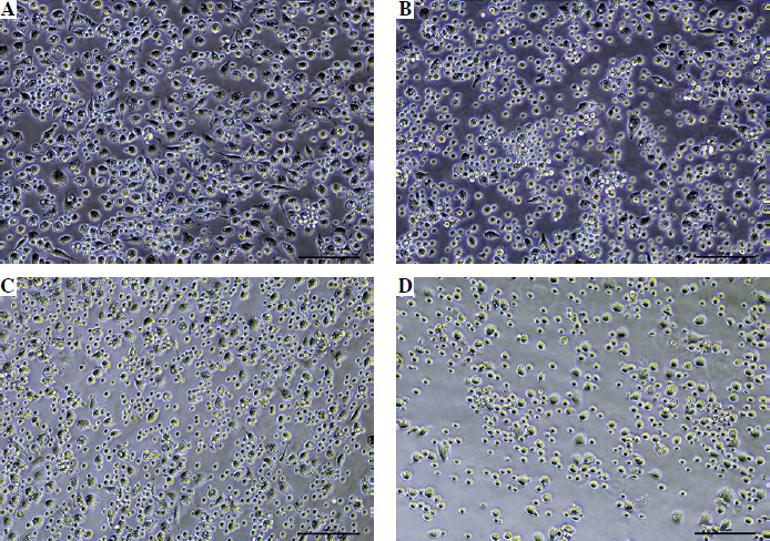
There was almost no difference in the morphology or quantity of the primary peritoneal macrophages between the MSC–/– and WT mice (Fig. 1A, C). However, at 24 hours after LPS stimulation, many of the cells in the MSC–/– group were shrunken and had poor viability compared with those in the WT group (Fig. 1B, D). Moreover, the number of primary macrophages in the MSC–/– group was lower than that in the WT group (Fig. 1B, D).
Fig. 1
Morphological differences in primary peritoneal macrophages among the different groups. A) WT control group, B) WT LPS group, C) MSC–/– control group, D) MSC–/– LPS group. The primary macrophages were cultured for 12 hours after adherence, after which LPS was added. After 24 hours, the morphological differences in the primary peritoneal macrophages in the different groups were observed
MSC deficiency influenced the adhesion function of the primary peritoneal macrophages
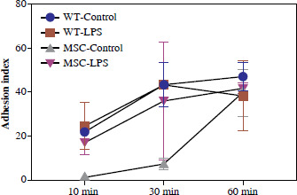
Compared with that in the WT group, the AI in the MSC–/– group was lower at 30 minutes. After LPS stimulation, the AI in the MSC–/– LPS group was still inferior to that in the WT LPS group. However, from 30 minutes to 60 minutes, the AI in the WT LPS group decreased, whereas that in the MSC–/– LPS group increased (Fig. 2).
Fig. 2
Dynamic changes in the AI in different groups. Primary peritoneal macrophages were cultured for 12 hours after adherence, after which LPS was added. After 24 hours, cells were collected in clean EP tubes and placed in a 37oC water bath. The cell suspensions were incubated in the water bath for 10, 30 and 60 minutes, and the AI was calculated according to the formula. MSC – MSC–/–
MSC deficiency inhibited the phagocytic function of the primary peritoneal macrophages after LPS stimulation
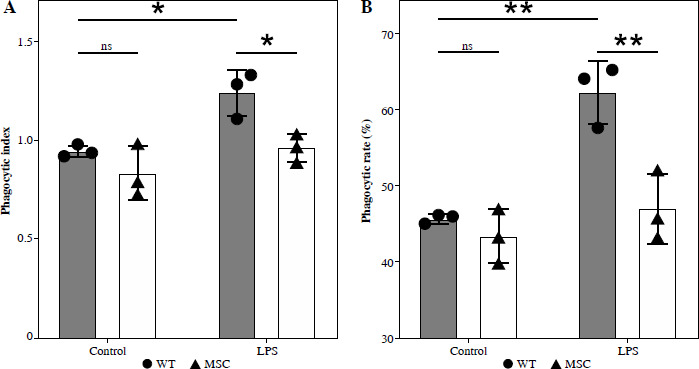
There was no significant difference in the phagocytotic ability of primary peritoneal macrophages between the MSC–/– control group and the WT control group (p > 0.05). However, after LPS stimulation, the number of E. coli phagocytosed by the primary peritoneal macrophages in the MSC–/– LPS group was significantly lower than that in the WT LPS group (Supplementary Fig. 1), while the number of E. coli phagocytosed by each cell was also reduced (Supplementary Fig. 1). Hence, the phagocytic index (Fig. 3A, p < 0.05) and the phagocytic rate (Fig. 3B, p < 0.01) results revealed that MSC deficiency (MSC–/–) inhibited the phagocytic function of the primary peritoneal macrophages.
Fig. 3
Phagocytic function of the primary peritoneal macrophages in different groups. Primary peritoneal macrophages were cultured for 12 hours after adherence, after which LPS was added. After 24 hours, E. coli were added to the cells at a cell : bacteria ratio of 1 : 10 in each group. Thirty minutes later, macrophage phagocytosis function was evaluated. A) Phagocytic index. B) Phagocytic rate. The respective significance levels are shown in the figure, significance level, *p < 0.05, **p < 0.01, ns – not significant, MSC – MSC–/–
MSC deficiency regulated the levels of inflammatory cytokines secreted by the primary peritoneal macrophages
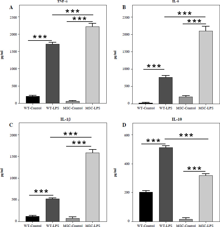
At 24 hours after LPS stimulation, the levels of proinflammatory and anti-inflammatory factors in the WT and MSC–/– groups were significantly higher than those in the control groups (p < 0.001, Fig. 4A-D). Moreover, the levels of proinflammatory cytokines, including TNF-α (Fig. 4A), IL-6 (Fig. 4B) and IL-1β (Fig. 4C), secreted by primary peritoneal macrophages in the MSC–/– LPS group were significantly higher than those in the WT LPS group (p < 0.001). In addition, compared with that in the WT group, the level of the anti-inflammatory cytokine IL-10 (Fig. 4D) in the LPS-stimulated group was significantly lower (p < 0.001).
Fig. 4
Levels of inflammatory cytokines secreted by the primary peritoneal macrophages in the different groups. Primary peritoneal macrophages were cultured for 12 hours after adherence, after which LPS was added. After 24 hours, the cell supernatants from each group were collected and the levels of inflammatory cytokines were detected. A) TNF-α, B) IL-6, C) IL-1β, D) IL-10. The respective significance levels are shown in the figure, ***p < 0.001. MSC – MSC–/–
MSC deficiency promoted the polarization of primary peritoneal macrophages towards the type 1 (M1) phenotype
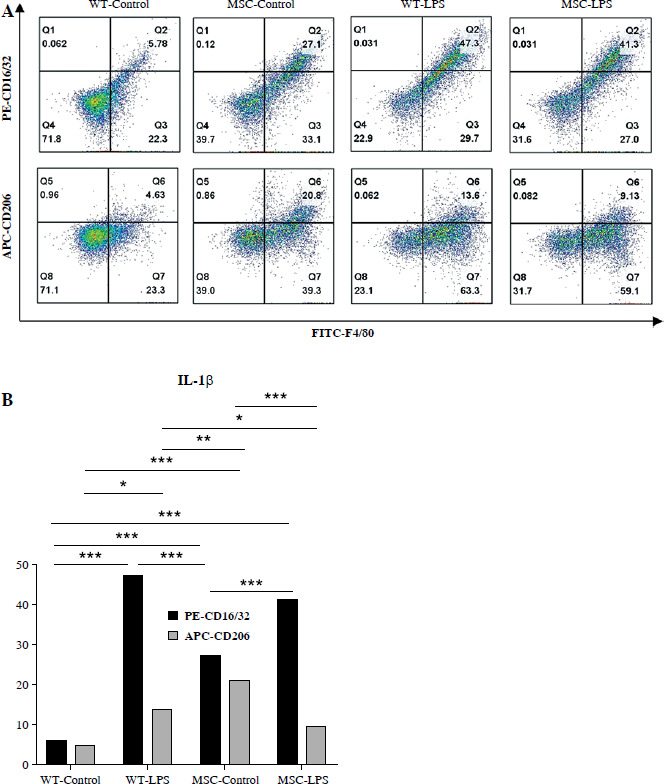
The numbers of M1 (CD16/32+) and M2 macrophages (CD206+) did not differ (5.78 vs 4.63) in the WT control group (Fig. 5A, B). Also, in the MSC–/– control group, the number of M1 (CD16/32+) macrophages and M2 macrophages (CD206+) showed the similar tendency (27.1 vs. 20.8) (Fig. 5A, B). However, the numbers of M1 and M2 cells in the MSC–/– control group were significantly greater than those in the WT control group (27.1 vs. 5.78; 20.8 vs. 4.63) (Fig. 5A, B). Compared with those in the WT control group, the numbers of M1 and M2 cells in the WT LPS group were higher (Fig. 5A, B). The proportion of M1 macrophages was increased in the MSC–/– group, whereas that of M2 macrophages was decreased (Fig. 5A, B).
Fig. 5
Polarization of primary peritoneal macrophages in different groups. Primary peritoneal macrophages were cultured for 12 hours after adherence, after which LPS was added. After 24 hours, cells were collected for staining for M1 (PE-CD16/32) and M2 (APC-CD206) markers and then subjected to flow cytometric analysis. A) Scatter plots. B) Bar charts. The respective significance levels are shown in the figure, *p < 0.05, **p < 0.01, ***p < 0.001. MSC – MSC–/–
Discussion
As important components of the immune system, macrophages play a key role in defending against pathogens, regulating inflammatory responses and promoting tissue repair [19]. Their regulatory functions influence the occurrence and development of infections. Recent research data have revealed that macrophages can act as immune modulators to enhance immune functions and improve the effectiveness of treatments that combat infections [20]. The main biological functions of macrophages, including adhesion, phagocytosis, secretion, and polarization, enable them to migrate to injured areas, clear pathogenic microorganisms and participate in inflammatory responses during infection [21]. Hence, studies on the regulatory factors of macrophages are helpful for improving immune regulation strategies aimed at preventing infection, and studying these factors has become an important research direction in the fields of inflammation, infection, and immunity.
MSC, which is expressed in multiple tissues and organs of mammals, is involved in the development, differentiation, and regeneration of skeletal muscle [9-15, 22]. In the past decade, many studies have shown that MSC is involved in the regulation of various immune cells. For example, MSC increases the expression level of Foxp3 by inhibiting the Th2 transcription programme and promotes the unidirectional differentiation of iTregs [16]. Moreover, MSC promotes the formation of memory B cells, prevents plasma cell differentiation, and ultimately affects the differentiation of B cells [23]. In addition, MSC deficiency affects the distribution of Tregs and causes a leftward shift in the Treg–Th17 balance after severe trauma [24]. These studies suggest that MSC in immunocytes is a potential regulatory target for the immune abnormalities observed in injury, disease, and infection. However, there have been no reports on the relationship between MSC and macrophages.
In this study, the primary peritoneal macrophages from WT and MSC–/– mice were collected, and LPS treatment was used to simulate bacterial infection ex vivo. The differences in the immune functions of the primary peritoneal macrophages in the different groups were subsequently explored. The results revealed that MSC had no obvious regulatory effect on the morphology or viability of primary peritoneal macrophages under normal conditions. However, under conditions simulating bacterial infection, MSC deficiency reduced the number of peritoneal primary macrophages and caused some of them to swell and undergo nuclear necrosis. These findings suggest that MSC may contribute to maintaining the structure and function of macrophages after infection.
After pathogens infect the host, macrophages can migrate to the lesion site and carry out their immune functions, and the strength of their adhesion ability can affect their recruitment and colonization efficiency [21, 25]. The results of this study revealed that MSC deficiency led to a decrease in the adhesion ability of primary peritoneal macrophages. Although LPS stimulation increased its adhesion ability within a certain time range, it was still lower than that in the WT LPS group. These findings suggest that MSC expression may be positively correlated with the adhesion ability of macrophages.
Macrophages regulate the inflammatory response by phagocytosing pathogens [26, 27] and secreting inflammatory cytokines [28-32]. The results of this study revealed that MSC deficiency reduced the phagocytic rate and phagocytic index of primary peritoneal macrophages compared with the WT LPS group after simulated infection. Moreover, the levels of pro-inflammatory cytokines, such as IL-1β, IL-6 and TNF-α, were higher, whereas the levels of anti-inflammatory cytokines, such as IL-10, were lower, relative to the WT LPS group. These data suggest that MSC may increase the phagocytic ability of macrophages and restore the balance between pro-inflammatory and anti-inflammatory responses after infection.
In addition, during infection, macrophages activate multiple signalling pathways, including the nuclear factor kappa-B (NF-κB), mitogen-activated protein kinase (MAPK) and phosphatidylinositol 3-kinase (PI3K) pathways, which promote the polarization of M1 macrophages and enhance the inflammatory response [33-37]. The results of this study showed that MSC deficiency reduced the proportion of M2 primary peritoneal macrophages but increased the proportion of M1 primary peritoneal macrophages compared with those in the MSC control group after simulated infection. These findings suggest that MSC may promote the polarization of macrophages from the M1 to the M2 phenotype and enhance their anti-inflammatory function. Notably, before LPS intervention, the numbers of M1 and M2 cells in the MSC–/– group were significantly higher than those in the WT control group, suggesting that the polarization of peritoneal macrophages was enhanced after MSC knockout. These findings also confirmed that MSC and macrophages are closely related in terms of function.
There are some limitations of this study. Only ex vivo experiments were performed, because the reproduction of MSC–/– mice is relatively difficult. Moreover, the primary peritoneal macrophages used in this study may not fully reflect the characteristics of all the macrophages. In addition, this study did not explore the molecular mechanism by which MSC affects macrophages. Therefore, it is necessary to verify these results using various types of macrophages and in vivo experiments in future studies. Additionally, the molecular mechanism by which MSC regulates the immune functions of macrophages will be further explored.
Conclusions
Taken together, the results of this study reveal the regulatory role of MSC in the immune functions of macrophages for the first time. MSC may enhance the immune function of peritoneal macrophages in mice, including reducing the adhesion index, increasing bacterial phagocytosis, decreasing the levels of proinflammatory cytokines, increasing the levels of anti-inflammatory cytokines, and shifting the polarization of macrophages from the M1 phenotype to the M2 phenotype. This study not only contributes to the understanding of how MSC regulates immunocytes but also provides a potential novel target of immune regulation for the prevention and treatment of infectious diseases.