Introduction
Cancer remains one of the leading causes of death worldwide, despite significant advances in traditional therapeutic approaches such as surgery, chemotherapy, and radiation therapy. These conventional treatments often fail to fully eradicate tumors, especially in the case of metastases or advanced-stage cancers, and they frequently come with severe side effects due to their lack of specificity. One of the most promising forms of cancer treatment in recent years is immunotherapy. Among the various types of immunotherapies, dendritic cell (DC)-based immunotherapy has gained particular attention due to the crucial role that DCs play in the initiation of adaptive immune responses [1]. DCs are highly effective in stimulating the immune system because of their unique ability to activate T cells, the central players in the immune response, by presenting tumor antigens through major histocompatibility complex (MHC) molecules [1, 2].
However, the effectiveness of DC-based immunotherapy is heavily dependent on both the number of DCs and the nature of the antigen presented to the immune system. Traditionally, DCs are differentiated from monocytes derived from peripheral blood [3, 4]. From 100 ml of peripheral blood, approximately 107 DCs can be obtained. During this process, it is generally believed that the number of DCs cannot be significantly expanded. The insufficient number of DCs severely limits their use in antigen-specific T cell proliferation, DC vaccine development, and the progress of related basic and clinical research [5].
Tumor-associated antigens (TAAs), which are expressed at higher levels on cancer cells compared to normal cells, serve as potential targets for anti-tumor immune responses. For DC-based immunotherapy to succeed, it is essential that the DCs are loaded with these specific tumor antigens, allowing them to effectively prime T cells to recognize and attack tumor cells. There are several strategies to load DCs with tumor antigens, including peptide-based loading, mRNA transfection, and protein-based methods [6]. Among these, the protein-based approach is particularly attractive because it allows for the presentation of naturally folded, immunologically relevant peptides to the DCs, which can then be processed and presented in a more physiologically relevant manner [7]. One promising protein for loading onto DCs is gp96, a highly conserved, multifunctional heat shock protein that serves as a molecular chaperone [8]. Gp96 can bind to the CD91 receptor on the surface of DCs, facilitating the internalization of the antigen into the DCs [9, 10]. Placental gp96, in particular, has been shown to bind a wide range of peptides, including those derived from tumor-associated antigens (e.g., GPC3, Muc-1, HER-2). As a molecular chaperone, placental gp96 aids in the proper folding of these peptides, ensuring that they are presented on the surface of DCs in a biologically active and immunogenic form [11]. This makes placental gp96 an ideal candidate for use in cancer immunotherapy, as it can present multiple tumor-associated peptides simultaneously. Consequently, placental gp96-loaded DCs are capable of activating T cells against a broad spectrum of tumor antigens, thus enhancing the likelihood of a successful immune response across different types of cancers [12, 13]. Furthermore, gp96 has been shown to be involved in the activation of both innate and adaptive immune responses, further enhancing its potential as a therapeutic agent.
In this study, we successfully expanded large quantities of mature DCs from peripheral blood precursors, and aimed to explore their potential when loaded with placental gp96 to generate antigen-specific T cells capable of recognizing and eliminating a wide range of tumor cells. Specifically, we focused on the ability of placental gp96-loaded expanded DCs to activate T cells in vitro, their efficiency in eliciting cytotoxic T lymphocyte (CTL) responses against various cancer cell lines, and their potential to target tumors with different expression profiles of gp96. Through this approach, we sought to establish the basis for developing a novel immunotherapeutic strategy that harnesses the power of the immune system to effectively target and eliminate tumor cells.
Material and methods
Expansion of dendritic cells from HSC
Peripheral blood (PB) samples were collected from 3 healthy donors after mobilization with granulocyte colony-stimulating factor (G-CSF, 10 µg/kg/day for 4-5 days). Mononuclear cells (MNCs) were isolated using Ficoll-Paque density gradient centrifugation, followed by two washes with phosphate-buffered saline (PBS). CD34+ hematopoietic progenitor cells were enriched using CD34+ magnetic-activated cell sorting (Stemery China) following the manufacturer’s protocol. The purified CD34+ cells were seeded in 24-well plates at a density of 5 × 10⁵/ml in RPMI 1640 medium supplemented with 10% FBS, 50 ng/ml granulocyte-macrophage colony-stimulating factor (GM-CSF, PeproTech USA), 50 ng/ml interleukin 4 (IL-4, PeproTech USA), and 100 ng/ml Flt3-ligand (FL, PeproTech USA). Cultures were maintained at 37°C in a humidified atmosphere with 5% CO2, and the medium was refreshed every two days by adding fresh cytokines. After 20-25 days of culture, immature dendritic cells (iDCs) were induced to mature by adding a maturation cocktail to the culture medium, consisting of 10 ng/ml lipo-polysaccharide (LPS, Sigma-Aldrich USA), 10 ng/ml tumor necrosis factor α (TNF-α, PeproTech USA), and 1 µg/ml CD40 ligand (CD40L, PeproTech USA). The cells were incubated for an additional 48 hours under the same culture conditions. Morphological changes indicative of maturation were observed under an inverted phase-contrast microscope. Dendritic cell phenotypes were analyzed using flow cytometry (Mindray China). Cells were stained with fluorophore-conjugated monoclonal antibodies specific for the following markers: CD80+, CD86+, CD83+, HLA-DR+ (BD Bioscience USA).
Generation of gp96-specific T cell lines
Immature DCs were incubated with 20 µg/ml of placental-derived gp96 (Heatshockbio China) for 24 hours to allow efficient loading. After incubation, the DCs were thoroughly washed with RPMI-1640 medium to remove unbound protein. The expression of surface markers, including CD80, CD86, and HLA-DR, was analyzed by flow cytometry (Mindray China) to confirm DC maturation. To generate antigen-specific T cells, naive T cells were isolated using the Naive CD8+T Cell Separation Kit (Stemery China) and subsequently co-cultured with DCs pulsed with placental gp96 at a DC : T cell ratio of 1 : 4. The culture was maintained in serum-free T cell medium (Stemery China) supplemented with 100 U/ml IL-2 (SL pharma China), 50 ng/ml IL-21, 100 ng/ml IL-7, and 20 ng/ml IL-15 (EastMab China). DCs were used to stimulate the T cells once a week for a total of four to five stimulations. Fresh culture medium was added every 4-5 days to maintain optimal conditions for T cell expansion.
T cell proliferation assessment
Cultured T cells were resuspended at 1-2 × 10⁶ cells/ml in RPMI-1640 with 10% FBS. CFSE was diluted (1-5 µM) in DMSO and added to the cell suspension, then incubated for 10-20 minutes at 37°C. After incubation, the reaction was quenched with RPMI-1640 medium and cells were washed twice by centrifugation (300 × g, 5 min). The labeled T cells were seeded at 1-2 × 10⁶ cells/ml in culture medium, and cultured with IL-2 (1000 U/ml) every 2-3 days. Cells were harvested at 3 days and analyzed for proliferation by flow cytometry.
NK cell expansion
Using the NK Cell robust Expansion Kit (Stemery China), high-purity NK cells were expanded from peripheral blood mononuclear cells. Briefly, mononuclear cells were isolated from collected peripheral blood using the Ficoll method. The cells were then seeded into anti-Her2 antibody coated cell culture flasks at a density of 1-2 × 10⁶/ml. Every 2-3 days, an equal volume of culture medium containing IL-2 (1000 U/ml) was added. Cells were harvested on day 14 of culture.
ELISPOT
A 96-well ELISPOT plate (Biosys Germany) was prepared and each well coated with 100 µl of anti-IFN-γ antibody diluted in PBS. The plate was incubated overnight at 4°C. The next day, the wells were blocked with 200 µl of complete RPMI-1640 medium for 2 hours at 37°C. Cultured T cells were collected and resuspended at 1-5 × 10⁵ cells/well in complete medium. 100 µl of the cell suspension was added to each well, and cells were stimulated with gp96 for 6-24 hours. The wells were washed, and biotinylated detection antibody (4A Biotech, China) was added for 2 hours, followed by streptavidin-HRP (Beyotime, China) for 1 hour. The substrate (AEC, Thermo Fisher, China) was applied, and the number of spots corresponded to the frequency of antigen-specific cytokine-producing cells.
Cytotoxicity assay
The human glioblastoma cell line U87MG (ATCC, USA) was used as a target cell line for cytotoxicity assays. T cells were co-cultured with U87MG cells at varying effector : target (E : T) ratios (1 : 1, 10 : 1) for 24 hours. Cytotoxicity was evaluated by 7-Aminoactinomycin D (7-AAD) (Beyotime China) flow cytometry assay. Cell viability was evaluated by 7-Aminoactinomycin D (7-AAD) calculated by comparing the survival of U87MG cells in the presence and absence of T cells. Effector cells (T cell) and target cells (U87MG) were co-incubated at different E : T ratios (1 : 1, 5 : 1, 10 : 1) in 24-well flat-bottom plates for 12 hours at 37oC in a humidified atmosphere containing 5% CO2. Target cells alone in culture medium were used as negative controls. After incubation, cell samples were collected and washed with ice-cold PBS once, then stained with 100 µl of 7-AAD for 5 minutes in the dark. The samples were analyzed on the flow cytometer. T cell cytotoxicity (%) was calculated as cells positive for 7-AAD/total CFSE-positive cells, after subtracting the spontaneous lysis (%) in the negative control. MHC restriction was assessed using blocking antibodies (10 µg/ml) against HLA-A2 (clone BB7.2; Neobioscience China) with mouse IgG isotype control, added at assay initiation.
Results
Generation of DC precursor cells
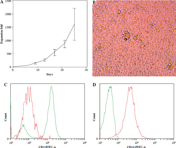
Hematopoietic stem cells derived from 3 healthy donors were expanded in vitro over a period of approximately 25 days. Compared to the starting cells, the final total cell number was amplified 942 to 2250 times (Fig. 1A). During the culture process, the cells gradually increased in size and became round (Fig. 1B), with a progressive decrease in the proportion of lymphocytes (Fig. 1C). These expanded cells served as precursor cells for DCs and were characterized by high expression of monocyte-specific marker CD14 (Fig. 1D).
Generation of gp96-pulsed mature dendritic cells
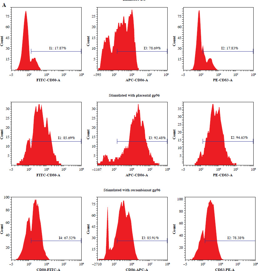
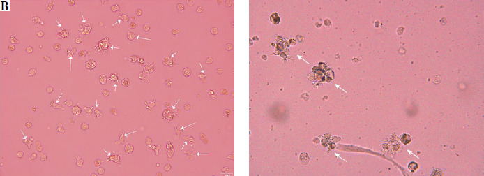
Under stimulation with GM-CSF and IL-4, DC precursor cells differentiated into immature DCs (iDCs). These iDCs exhibited moderate expression of CD80, CD83, CD86, and HLA-DR proteins. Subsequently, the iDCs were exposed to placental gp96 protein, and upon stimulation with DC maturation-inducing factors such as LPS, TNF-α, and CD40L, the iDCs underwent differentiation into mature dendritic cells. The resulting mature dendritic cells displayed typical phenotypic features of mature DCs, including high expression of markers such as CD80, CD83, and CD86 (Fig. 2A). Additionally, the use of recombinant gp96 to sensitize DCs also facilitated the generation of mature dendritic cells (Fig. 2A). Finally, the generated cells exhibited the typical morphological characteristics of DCs (Fig. 2B).
T cell proliferation
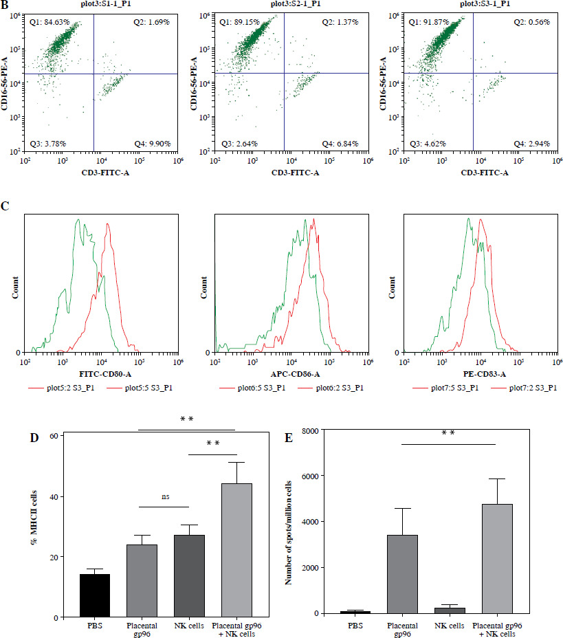
After four to five rounds of weekly stimulation with placental gp96-loaded DCs, significant proliferation of T cells was observed. Compared to the control group (unmodified DCs), where T cells did not proliferate, T cells stimulated by DCs loaded with placental gp96 exhibited three CFSE peaks, indicating that the cells had undergone at least two divisions (p < 0.001). These results suggest that placental gp96-loaded DCs effectively activated naive T cells. However, T cell proliferation was also observed in DCs pulsed with recombinant gp96 (Fig. 3A). Additionally, NK cells were included to validate DC-NK crosstalk in enhancing DC maturation. We generated high-purity NK cells in vitro using PBMCs from the same donor that was used for generating the DCs (Fig. 3B). When NK cells were co-cultured with gp96 and T cells, it was found that NK cells facilitated the gp96-induced maturation of DCs, resulting in elevated expression of various surface molecules associated with MHC II, CD80, CD83 and CD86 (Fig. 3C, D). DCs co-cultured with NK cells and placental gp96, under the same stimulation conditions and at equivalent frequencies, were able to generate a higher number of antigen-specific T cells (Fig. 3E).
Fig. 3
T cell activation and proliferation. A) Proliferation of T cells upon stimulation by different types of dendritic cells (unmodified DC: expanded DCs without protein loading were used to stimulate T cells)
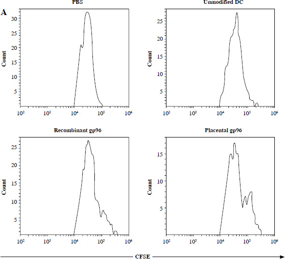
Fig. 3. Cont.
B) In vitro cultivation of NK cells derived from peripheral blood mononuclear cells (PBMCs); C) Impact of NK cells on maturation of placental gp96 protein-pulsed DCs; D) Effect of NK cells on expression of MHC class II molecules on placental gp96 protein-pulsed DCs; E) Influence of NK cells on generation of antigen-specific T cells from placental gp96 protein-pulsed DCs
Cytotoxicity against tumor cell lines
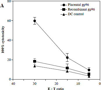
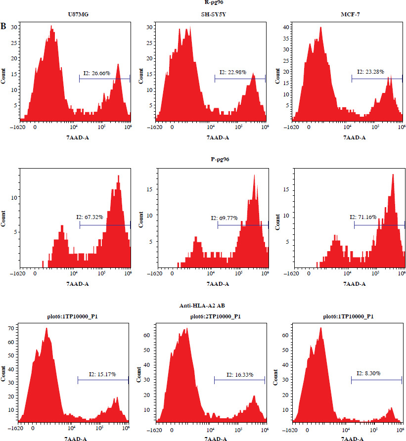
The cytotoxic activity of placental gp96-activated T cells was evaluated. Initially, we co-cultured the generated T cells with placental gp96-pulsed DCs and observed that the T cells effectively lysed placental gp96-pulsed DCs. In contrast, their cytotoxicity against recombinant gp96-pulsed DCs or unmodified DCs was significantly reduced (Fig. 4A). To assess the cytotoxicity of T cells generated by placental gp96-pulsed DCs against tumor cells, we selected the glioblastoma cell line U87MG, the neuroblastoma cell line SH-SY5Y, and the breast cancer cell line MCF-7. Both the T cell donor and tumor cell lines were HLA-0201 positive. At an effector : target (E : T) ratio of 10 : 1, T cells exposed to placental gp96-loaded DCs demonstrated approximately 67.32% cytotoxicity against U87MG glioblastoma cells, 69.77% cytotoxicity against SH-SY5Y neuroblastoma cells, and up to 71.16% killing efficiency against MCF-7 (Fig. 4B). In contrast, T cells co-cultured with unmodified DCs exhibited less than 30% cytotoxicity at the same ratio. Furthermore, T cells generated from recombinant gp96-pulsed DCs displayed tumor cell killing efficiencies of 26.66%, 22.98%, and 23.28% against the three tumor cell lines, respectively, which were notably lower than the cytotoxicity observed for T cells generated by placental gp96 pulsed DCs (Fig. 4B). Additionally, T cells generated from HLA-0201 donors did not exhibit cytotoxicity against non-HLA-0201 tumor cell lines. To determine MHC restriction of these T cell lines, we examined the inhibitory effects of anti-MHC antibodies on their cytolytic activity. Following HLA-A2 antibody blockade, the killing efficacy of gp96-specific T cells against U87MG, SH-SY5Y, and MCF-7 cells was significantly reduced by 52%, 53%, and 63%, respectively (p < 0.01), providing additional evidence of the HLA restriction of their killing activity (Fig. 4B). These findings suggest that placental gp96-loaded DCs effectively stimulate T cells to recognize and eliminate U87MG glioblastoma cells, SH-SY5Y neuroblastoma cells, and MCF-7 breast cancer cells, with this cytotoxicity being HLA-restricted.
Discussion
In this study, we aimed to evaluate the potential of placental gp96-loaded dendritic cells (DCs) in generating antigen-specific T cells capable of inducing potent anti-tumor immunity. The cytotoxicity assays revealed that T cells generated through exposure to placental gp96-loaded DCs exhibited substantial cytotoxicity against multiple tumor cell lines.
Traditionally, DCs have been considered difficult to expand efficiently in vitro, which has posed a significant challenge for advancing research and clinical applications. In response to this limitation, several research groups have explored methods for the ex vivo expansion of DCs, yielding promising results [14-16]. Notably, some studies have successfully expanded large populations of DCs from umbilical cord blood CD34+ cells [17]. Building upon this foundation, our team has optimized these protocols and achieved successful expansion of DCs from peripheral blood CD34+ cells. Furthermore, with continued refinement, we have developed a method that enables the efficient expansion of DCs directly from mononuclear cells (MNCs) without the need for prior selection of CD34+ cells (data not shown). These advances represent a significant step forward in addressing the challenge of insufficient DC numbers in clinical applications, offering a robust approach for large-scale DC production for therapeutic use.
As is well known, the placenta and tumors share many similarities [18]. The concept of using placental-derived components for cancer immunotherapy is based on the biological similarities between the placenta and tumors, particularly in their shared mechanisms of immune modulation and immune evasion. Both tissues, while playing very different roles in the body, use similar strategies to avoid immune detection. These similarities make the placenta an intriguing model for designing cancer immunotherapies that could effectively target and eliminate tumor cells.
T cells generated from expanded DCs pulsed with placental gp96 exhibit stronger anti-tumor effects compared to those generated from recombinant gp96-pulsed DCs. This may be attributed to the presence of a wide variety of natural tumor-associated peptides on placental gp96. Placental gp96 is a versatile carrier of tumor-associated antigens. It is capable of binding a broad array of peptides, including those derived from well-known tumor antigens (e.g., HER2, GPC3), as well as novel or uncharacterized tumor-specific antigens [11, 13]. This ability enables it to target a wide spectrum of cancers, offering the potential for personalized immunotherapy tailored to the unique antigenic profile of each patient’s tumor. The recombinant gp96 protein does not carry any tumor antigens, which may explain the reduced anti-tumor capability of T cells generated by DCs loaded with recombinant gp96. However, recombinant gp96 itself or after modification may possess favorable tumor antigen-binding capacity [19]. Further studies are needed to investigate whether antigen-specific T cells generated by DCs loaded with tumor antigen-bound recombinant gp96 exhibit tumor-killing activity. This approach may provide an alternative solution to address the limited availability of natural gp96 proteins (both placental-derived and tumor tissue-derived).
T cells generated from expanded DCs pulsed with placental gp96 exhibit potent tumor cell-killing activity in vitro. Future studies should focus on optimizing this approach for clinical applications, including evaluating its efficacy in animal models and exploring its potential to be combined with other immunotherapies, including potential combination strategies with immune checkpoint inhibitors (e.g., anti-PD-1/PD-L1 antibodies) to enhance therapeutic efficacy. Additionally, the applicability of this method to various cancer types and the underlying mechanisms of T cell-mediated killing in vivo require further investigation.
Conclusions
This study demonstrated that placental gp96 can effectively generate a large number of antigen-specific T cells in vitro with anti-tumor activity. This activity may be mediated by various tumor-associated peptides presented on the placental gp96. Moreover, this anti-tumor effect is MHC-restricted. These findings suggest that placental gp96-loaded DCs hold significant promise as a novel immunotherapy strategy for cancers.